The mitral valve sits between 2 chambers of the heart called the left atrium (that receives oxygenated blood from the lungs) and the left ventricle (the main pumping chamber of the heart). The valve leaflets are held together by intricate apparatus consisting of small tendons and muscles. Damage to any part of this can cause the valve to become leaky and the flow of blood is reversed from the left ventricle into the left atrium. This is called mitral regurgitation. If the mitral valve becomes narrowed, preventing blood from entering the left ventricle, is it called mitral stenosis.
Mitral regurgitation
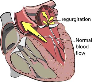
Mitral regurgitation is the most common form of valvular heart disease. Mitral valve prolapse is most frequently the cause of regurgitation. It is found typically in young females and as one gets older, the valve leaflets degenerate. Ischaemic heart disease is another cause of mitral regurgitation, caused by damage to the valve apparatus and small muscles that hold the valve leaflets together. Rarer causes include: connective tissue disorders, e.g. Marfan's, rheumatic fever, endocarditis and cardiomyopathies. The following diagram illustrates the regurgitation across the mitral valve instead of following the normal blood flow pattern.

Breathlessness and palpitations are a common presenting symptom in severe cases as patients are more prone to heart failure and resulting fluid congestion in the lungs. However, many patients with mild or moderate mitral regurgitation may remain asymptomatic.
An ECG will help detect any rhythm abnormalities such as atrial fibrillation, an irregular heart beat that is commonly associated with mitral valve disease. An echocardiogram will best visualise the mitral valve anatomy and assess the severity of the regurgitation. A chest X-ray will also help detect any fluid on the lungs in more severe cases.
Medical therapy to help alleviate the strain on the heart is the first-line therapy. In severe mitral regurgitation, when the left ventricle is showing signs of stretching, mitral valve repair or replacement may be required.
Mitral stenosis
Mitral stenosis is when the mitral valve becomes narrowed and stiffer, resisting the blood flow into the left ventricle which is the main pumping chamber of the heart.
The most common cause of mitral stenosis is previous rheumatic fever. The advent of antibiotic treatment has reduced the prevalence of rheumatic fever and subsequently, the number of patients with mitral stenosis has also fallen. Rarer causes are congenital calcification of the mitral valve or previous endocarditis.
Heart failure symptoms such as breathlessness, especially on exertion are common. Patients may also feel palpitations and chest pain. If the heart rate is irregular (atrial fibrillation), clots may form in the heart that can be swept away in the blood to and block smaller arteries further upstream. This can result in a stroke.
An ECG will help detect any underlying rhythm disturbances such as atrial fibrillation. An echocardiogram will diagnose the severity of the mitral stenosis and also its effect on the heart muscle and lung pressures. A more accurate way of determining lung pressures is by cardiac catheterization.
Again, treatment focuses initially on symptom control and removing excess fluid from the lungs through medication. If there are severe clinical symptoms and echocardiographic evidence, a mitral valve repair, replacement or valvuloplasty may be suitable. This will be determined by your cardiologist.
Contact Us
01494 867 616
info@chilternheart.com
To book an appointment click here
